Shoulder Bone Anatomy Diagram / Anatomy Of The Human Shoulder Joint. A fractured clavicle is the most frequently broken bone in the body. The human shoulder is made up of three bones: This image added by admin. 8 name the arteries and the nerves that supply shoulder joint. We think this is the most useful anatomy picture that you need.
The shoulder is composed of a network of bones, joints, and soft tissues that make this large range of motion possible. All about the shoulder muscles. We think this is the most useful anatomy picture that you need. Four rotator cuff muscles that act on the shoulder begin at the scapula. A fractured clavicle is the most frequently broken bone in the body.
Arm And Shoulder Anatomy Bones Muscles And Nerves Kenhub from thumbor.kenhub.com Shoulder joint of human body anatomy infographic diagram with all parts including bones ligaments muscles bursa cavity capsule cartilage membrane for medical. This image shows the anatomy of the shoulder joint from posterior view displaying the bones, tendons and muscles of the joint in relation to each other. Attaching to the clavicle are the pectoralis major, sternocleidomastoid, deltoid and trapezius muscles. Four rotator cuff muscles that act on the shoulder begin at the scapula. All about the shoulder muscles. It is divided into the head, anatomic neck, surgical neck, and shaft. Simple structure of the clavicle. Anchors for the torn tendons, ac shaving , biceps tendon groove shaving , etc.
We think this is the most useful anatomy picture that you need.
The shoulder is not a single joint, but a complex arrangement of bones, ligaments, muscles, and tendons that is better called the shoulder girdle. Related online courses on physioplus. The human shoulder is made up of three bones: Anchors for the torn tendons, ac shaving , biceps tendon groove shaving , etc. Home > blog > anatomy > shoulder anatomy: Very soon we'll move on to muscles! Acromion anatomy, ankle bones anatomy, bones of the clavicle, elbow bones anatomy, hip bones anatomy, shoulder muscles anatomy, shoulder pain anatomy, hand, acromion anatomy, ankle bones anatomy related posts of shoulder bones anatomy diagram. Click and start learning now! The bones of the shoulder consist of the humerus (the upper arm bone), the scapula (the shoulder blade), and the clavicle (the collar bone). The shoulder is composed of a network of bones, joints, and soft tissues that make this large range of motion possible. You can click the image to magnify if you cannot see clearly. The head of the humerus forms the ball. We think this is the most useful anatomy picture that you need.
Home > blog > anatomy > shoulder anatomy: The bones of the shoulder consist of the humerus (the upper arm bone), the scapula (the shoulder blade), and the clavicle (the collar bone). Normal anatomy, variants and checklist. This image added by admin. The shoulder joint has the largest range of motion out of all the joints in the body.
Anatomy 101 Shoulder Bones The Handcare Blog from blog.handcare.org Very soon we'll move on to muscles! Home > blog > anatomy > shoulder anatomy: Acromion process of the scapula 5. The shoulder anatomy includes the anterior deltoid, lateral deltoid, posterior deltoid, as well as the 4 then there was the bone work; Shoulder anatomy, shoulder bone, shoulder diagram, shoulder joint bones, shoulder muscle structure, shoulder parts of the body, shoulder pectoral girdle anatomy bones muscles function diagram ehealthstar shoulder anatomy images stock photos vectors shutterstock human skeletal. You can click the image to magnify if you cannot see clearly. Click now to learn about the bones, muscles, arteries, and nerves of these regions! 8 name the arteries and the nerves that supply shoulder joint.
The shoulder joint has the largest range of motion out of all the joints in the body.
Related online courses on physioplus. Shoulder anatomy, shoulder bone, shoulder diagram, shoulder joint bones, shoulder muscle structure, shoulder parts of the body, shoulder pectoral girdle anatomy bones muscles function diagram ehealthstar shoulder anatomy images stock photos vectors shutterstock human skeletal. All about the shoulder muscles. The shoulder joint is the connection between the chest and the upper extremity. The bones of the shoulder consist of the humerus (the upper arm bone), the scapula (the shoulder blade), and the clavicle (the collar bone). This image added by admin. Robin smithuis and henk jan van der woude. Click and start learning now! Shoulder bone on white background. The deepest layer of the shoulder includes the bones and the joints. Capitulum is specialized portion of hinge joint that allows radial head rotation. Simple structure of the clavicle. Spine of the scapula 3.
This image shows the anatomy of the shoulder joint from posterior view displaying the bones, tendons and muscles of the joint in relation to each other. We think this is the most useful anatomy picture that you need. Anatomynote.com found shoulder bone anatomy from plenty of anatomical pictures on the internet. The head of the humerus forms the ball. The deepest layer of the shoulder includes the bones and the joints.
Shoulder Physiopedia from www.physio-pedia.com This image added by admin. Master arm and shoulder anatomy by studying this topic page at kenhub. The shoulder is not a single joint, but a complex arrangement of bones, ligaments, muscles, and tendons that is better called the shoulder girdle. Acromion anatomy, ankle bones anatomy, bones of the clavicle, elbow bones anatomy, hip bones anatomy, shoulder muscles anatomy, shoulder pain anatomy, hand, acromion anatomy, ankle bones anatomy related posts of shoulder bones anatomy diagram. Attaching to the clavicle are the pectoralis major, sternocleidomastoid, deltoid and trapezius muscles. Long bones, short bones, and flat bones. The next layer is made up of the ligaments of the joint capsule. The shoulder is composed of a network of bones, joints, and soft tissues that make this large range of motion possible.
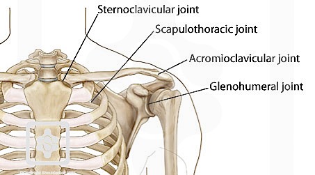
The clavicle (collarbone), the scapula (shoulder blade), and the humerus (upper arm bone) as well as associated muscles, ligaments and tendons.
Normal anatomy, variants and checklist. Learn vocabulary, terms and more with flashcards, games and other study tools. Spine of the scapula 3. The clavicle and scapula form the shoulder girdle. The shoulder joint has the largest range of motion out of all the joints in the body. The head of the humerus forms the ball. A fractured clavicle is the most frequently broken bone in the body. The human shoulder is made up of three bones: Click and start learning now! Related online courses on physioplus. Anchors for the torn tendons, ac shaving , biceps tendon groove shaving , etc. Editor · aug 6, 2017 ·. Shoulder joint of human body anatomy infographic diagram with all parts including bones ligaments muscles bursa cavity capsule cartilage membrane for medical.
The bones of the shoulder consist of the humerus (the upper arm bone), the scapula (the shoulder blade), and the clavicle (the collar bone) shoulder anatomy diagram. Learn vocabulary, terms and more with flashcards, games and other study tools.
Share :
Post a Comment
for "Shoulder Bone Anatomy Diagram / Anatomy Of The Human Shoulder Joint"


:background_color(FFFFFF):format(jpeg)/images/library/10813/image4.png&description=Shoulder Bone Anatomy Diagram / Anatomy Of The Human Shoulder Joint)
Post a Comment for "Shoulder Bone Anatomy Diagram / Anatomy Of The Human Shoulder Joint"